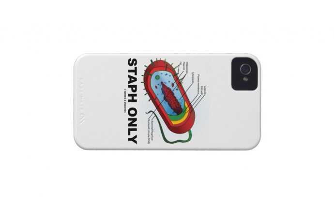
Bacterium Diagram
The illustration shows a generalised bacterium with many of the main components illustrated. No real organism would have all of these features. The image above is 400 pixels across and the original is 5, 438 pixels across.
CELL WALL
The top half of the diagram shows a Gram +ve bacterium with its thick peptidoglycan outer wall closely apposed to the inner plasma membrane. The lower half of the cell shows a Gram -ve bacterium with its inner plasma membrane, thin intermediate peptidoglycan layer and external membrane. The outer portion of the external membrane is a lipopolysaccharide layer. This layer comprises core components that form the outer layer of the membrane and (usually) side chains that radiate off. These side chains are called O-antigens. They are absent in Yersinia pestis. In this diagram, parts of the two types of wall are covered in a regular arrangment of proteins called an S-layer. A good example of an S-layer is shown in our diagram of anthrax.
CAPSULE
A generalised capsule is shown covering part of the Gram +ve and Gram -ve regions. Capsules can prevent drying, help in adhesion and help to ward off attack from viruses and host cells.
SURFACE FEATURES
Several flagella are shown. These are spiralised protein tubes that have a motor at their base. This motor anchors the flagella into the cell wall and its rotation causes the flagella to propel the bacterium along, like the propeller of a boat. Various flagellar arrangements are possible, from a single flagellum at one pole to numerous flagella radiating from all over the bacterial surface. Straighter protein tubes called pili are also shown. These can help in attachment of bacteria to host cells or other substrates. Pili are also used for gene transfer.
GENOME
The genetic material is a skein of circular DNA localised as the nucleoid. The nucleoid lacks a nuclear membrane (a defining characteristic of prokaryotic cells). Peeping out from the upper right part of the nucleoid is a plasmid - a separate piece of DNA. Plasmids replicate independently of the nucleoid DNA and can be exchanged between cells (the means of passing on, for example, drug resistance).
CYTOPLASM
The bacterial cytoplasm is shown filled with ribosomes. These are somewhat smaller than their eukaryotic counterparts and many are shown linked into polysomes. Infoldings of the Plasma membrane are common and are often associated with the nucleoid. In this picture such an infolding can be seen around the middle of the cell and it is termed a mesosome.
HELICAL FILAMENTS
Helically arranged protein filaments are shown winding around just beneath the plasma membrane. They are thought to guide the production of the cell wall to create the shape of the bacterium. They are absent in spherical forms. The purple helix (MreB) controls cell width and the bluish one (Mbl) controls length. They are composed of actin like molecules. This form of prokaryotic cytoskeleton was recently described in the journals CELL and NATURE (references: CELL, Vol. 104, 913-922, March 23, 2001. Nature, Vol. 413, September 2001).


